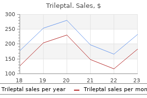
Trileptal dosages: 600 mg, 300 mg, 150 mg
Trileptal packs: 30 pills, 60 pills, 90 pills, 120 pills, 180 pills, 270 pills, 360 pills

Binary vectors are based on plasmids stable in both Escherichia coli and Agrobacterium medicine ethics cheap 600 mg trileptal free shipping. Because cointegrative vectors are based on the Ti plasmid, they are very stable in Agrobacterium, but they can be difficult to work with in practice. On the other hand, binary vectors can be handled with relative ease, although they are unstable in Agrobacterium and continued antibiotic resistance selection is required to maintain them (1). Transformation using Agrobacterium requires direct contact between the plant cell and the infecting bacteria. In practice, this is achieved by cocultivation of Agrobacterium with isolated plant cells or explants, such as leaves, roots, or tubers. In this case, bacteria are incubated with plant material, and transformation occurs over a period of about two days. Then, the plant material is washed to remove excess bacteria and cultured further to allow plant regeneration (1). Recently, a novel nontissue culture approach of transformation has been described using the model plant Arabidopsis. Here the developing floral buds of intact plants are vacuum infiltrated with the Agrobacterium. It has been generally assumed that Agrobacterium-mediated transformation is limited to the obvious natural hosts of Agrobacterium, the dicotyledonous plants. Recently, however, it has been established that Agrobacterium can be used effectively to create transgenic monocotyledonous plants, such as rice (3). The only requirement is that the plasmid contains a gene linked to a plant-specific promoter that is functional in transgenic plants. The two techniques used most frequently with isolated cells, treatment with polyethylene glycol and electric discharge or electroporation (see Transfection), achieve relatively high frequencies of transformation, which generally result in measurable activity of the transferred gene constructs after approximately one day. This allows transient expression assays, for example, to test the expression of specific promoter/gene constructs. Cells transformed in this manner can also be regenerated to intact plants as described previously. Upon insertion into the genome, a gene linked to a plant-specific promoter is expressed in a pattern determined by that promoter.
Farr (1967) "The ammonium sulphate method to measure antigen binding capacity" medications given during dialysis purchase trileptal 150 mg on line, in Handbook of Experimental Immunology, D. Classical Fate Maps A fate map is a graphic representation of the cells or regions within the embryo at one point during development that will develop into specific tissues and organs at subsequent developmental stages. The concept of the fate map may be one of the earliest abstract ideas in embryology, one that is implicit, for example, in the "homunculus" imagined by seventeenth century microscopists to be a miniature adult human that is "preformed" within the sperm. The concept of preformation was generally favored by scientists until the last half of the eighteenth century, when Caspar Friedrich Wolff, through observation of chick embryo development, postulated that embryos develop through a process he termed "epigenesis," whereby unpatterned tissues of the early embryo reorganize and expand to form the tissues and organs of the adult. As the cellular basis for tissue organization came to be understood by the mid-nineteenth century, it eventually became clear that embryonic development could be characterized by four cellular phases common to all vertebrate and most invertebrate embryos: (i) formation of the single-celled, diploid zygote (or fertilized egg) through union of haploid sperm and egg; (ii) cleavage, in which the fertilized egg undergoes mitotic division without increase in size to form the multicellular blastula (mammalian: blastocyst); (iii) gastrulation, in which blastula cells rearrange and expand to form the three primary germ layers (ectoderm, mesoderm, and endoderm) that are organized into the basic body plan (concepts developed in the early nineteenth century by von Baer); and (iv) organogenesis, in which the cells within the the three primary germ layers of the embryo form the organ and tissue primordia that develop into the tissues and organs of the fetus. The fetal stage of development is distinguished from the embryonic phase in that the fetus is indeed a preformed miniature of the adult that contains all of the organ and tissue rudiments (in the right places and containing differentiated cells and stem cells) that need only to expand and mature to function as adult tissues and organs. The transition from embryo to fetus occurs at about the eighth week of human development. In the late nineteenth and early twentieth centuries, embryologists began to take an experimental approach to the study of development. Fate maps of relatively simple embryos, such as ascidians, were initially established by directly observing the fate of blastula cells during gastrulation (1). The emergence of dyes that could be applied to living tissues without damaging them ("vital dyes") permitted selective staining of parts of blastula embryos and subsequent tracing of the tissues of the embryo that developed from these stained parts. Systematic application of this procedure to trace the fate of different regions of amphibian blastulas through gastrulation led to the construction of the first detailed and comprehensive fate map of a vertebrate embryo (2, 3). Subsequent work by many individuals led to the construction of fate maps for organ rudiment primordia of a variety of chordate embryos (amphioxus, fish, amphibians, birds) (4-9). These maps continue today as the primary basis for our present day understanding of morphogenetic movements (2) that blastula cells undergo during gastrulation. Nevertheless, these maps are constantly being revised and refined by applying more modern techniques. Although fate maps, such as those described previously, give a general idea how regions of the blastula give rise to the primary organ primordia of the embryo, they do not yield precise details of the way cells within those primordia give rise to the more complex tissues and organs of the fetus and adult.
Syndromes
Cleavage of the substrate occurs at the glycosidic bond between the residues bound to the D and E sites medications high blood pressure trileptal 600 mg purchase free shipping. The sugar ring at D site must be distorted from the normal chair conformation to a half-chair form to achieve good contacts with the enzyme. Lysozyme residues Glu35 and Asp52 are in position near the cleavage site and are considered to be the primary catalytic residues. Glu35, which lies in a hydrophobic environment, participates in catalysis in its protonated form, while Asp52, which is in a hydrophilic environment, does so in its nonprotonated form. As shown in Figure 3, Glu35 donates its proton to the oxygen linking the D and E sugars. A carbonium ion is formed on the D sugar, and this ion is stabilized by Asp52 and by the formation of an oxocarbonium ion. Formation of the planar oxocarbonium ion is favored by the distortion of the D sugar. As a result of this stabilization of the transition state, lysozyme can rapidly hydrolyze glycosidic bonds (4). This is a good example of how enzymes generally stabilize the transition state of their enzymatic reaction (see Catalysis). The reaction is completed by one-half of the cleaved product dissociating, and water replacing it and reacting with the bound intermediate. Extraneous oligosaccharides can also bind to the vacated sites and can compete with water for the last step. Physiological Function Lysozyme is widely distributed in nature, so some biological functions (8) other than its primary anti-bacterial action are expected. Lysozyme is effective against bacterial and viral infections and also shows antiphlogistic activity in a number of pathological conditions. Owing to these effects, lysozyme is employed as a medicine and as a food additive. Lysozymes are used as digestive enzymes in some organisms, such as ruminants and insects. Comparative Biochemistry Lysozymes are widely distributed in nature, from humans to viruses and plants.

As a consequence medications memory loss buy trileptal 600 mg low cost, the enzyme is stabilized in a form in which the active site, located at a distance from the phosphorylated serine, becomes more accessible to the substrate. In this case, the effects of phosphorylation on the structure of the enzyme are comparable to those occurring during classical allosteric activation, except that the effects of phosphorylation are due to a covalent modification of the enzyme, and not to ligand binding. Effects of Phosphorylation on ProteinProtein Interactions Protein phosphorylation can have dramatic regulatory effects on proteinprotein interactions, as disclosed by the study of protein tyrosine phosphorylation. Such enzymes, including phosphatidylinositol-3-kinase, phospholipase Cg, or guanine-nucleotide exchange factors, are thus brought into the vicinity of their membrane-associated substrates. The Place of Protein Phosphorylation in the Signal Transduction Networks Many signal transduction systems in cells use several modules whose hierarchical organization allows powerful amplification and precise modulation. Protein phosphorylation is involved in most of these transduction systems, either as an output mechanism, by altering the properties of target proteins, or as a module of information processing. Indeed, phosphorylation reactions in cells often occur in cascades in which activation of a first protein kinase phosphorylates and activates a second protein kinase, which, in turn, phosphorylates and activates a third kinase, and so on. This situation may account in part for the very large number of protein kinases, and it apparently has a number of interesting properties (40, 41). First, phosphorylation cascades allow amplification of the signal: One molecule of the first activated kinase can phosphorylate several secondary kinases, which can each phosphorylate several tertiary kinases, and so on. Another property of these cascades is to acquire kinetic properties, such as positive or negative cooperativity, similar to those of allosteric enzymes, even if the kinases composing the cascade themselves follow classical MichaelisMenten kinetics. Thus, protein kinase cascades can convert graded inputs into switch-like outputs (42). Finally, the kinase cascades provide many levels of regulation and possible cross-talks between different regulatory pathways. Thus, it is likely that the organization of protein kinases in cascades confers strong evolutionary advantages, as attested by the very high degree of conservation of many of these cascades. Examples of Phosphorylation Dysfunction in Human Diseases As mentioned above, virtually all intracellular biological processes are regulated to some extent by protein phosphorylation.
In that sense treatment with chemicals or drugs 150 mg trileptal order mastercard, they differ from hydrophobicity, which involves a second solvent that is immiscible with water, and with which the solute molecule interacts more or less strongly (see Partition Coefficient). Hydrophilic character also differs from solubility, in which the reference phase is the pure solute in liquid or crystalline form: Such an arbitrary reference is of questionable value if different solute molecules are to be compared against each other. Hydrophilic character can be determined in some cases by measuring the water solubility of a gas under an atmosphere of known partial pressure, or conversely, by measuring the concentration of a solute in the gas space over an aqueous solution of known concentration. In more typical cases, of the kind often encountered in biological molecules, the concentration of gas in the vapor phase may be too low to detect directly. In such cases, a large volume of carrier gas (usually argon or nitrogen) is bubbled through an aqueous solution of known concentration, then through an efficient trap, which may be water itself. From the amount of solute collecting in the trap, plotted as a function of the measured volume of carrier gas that has passed through the system, it is a simple matter to calculate the concentration of solute in the vapor phase. A scale of such equilibrium constants for simple organic compounds in uncharged form is shown in Figure 2. From the behavior of methylamine and acetic acid, eg, it can be inferred that the concentration of glycine in the vapor phase, over a 1- M aqueous solution, is approximately 1014M, even though that value is too low to measure directly. The equilibrium for transfer of a molecule from dilute aqueous solution to the vapor phase. Hydrophilicity is the reciprocal of this equilibrium constant, and it measures the affinity of a molecule for watery surroundings. These methods have revealed that the hydrophilicities of the protein amino acid side-chains, expressed in terms of their equilibria of transfer from the vapor phase to neutral aqueous solution at 25°C, span a range of approximately 1016-fold(3, 5). Figure 3 shows the relationship between hydrophilicity and the nominal transfer free energy of each side-chain from the interior to the surface of a protein, as indicated by the relative populations of surface and buried residues for each amino acid. In addition, a remarkable bias in the genetic code was noted, such that purines (usually A) serve as the second code letter for all the more hydrophilic amino acids (3, 4). Figure 4 shows the amino acid side-chains in order of increasing hydrophilicity, alongside the code letters at the second position for each of the amino acids, but the significance of this correlation is not understood. Tendencies of the 20 amino-acid residues to appear at the surfaces of globular proteins, compared with their free energies of transfer from the vapor phase to neutral aqueous solution (3, 4). Code letters and anticode letters at the second position for each of the amino acids.
Rasarus, 54 years: In that case, a D-J-Cm protein is synthesized, which becomes exposed at the surface of the preB cell and blocks any further gene rearrangement (see B Cell), but is unable to contribute a functional immunoglobulin.
Jared, 46 years: Green Fluorescent Protein Aequorea (and some other bioluminescent coelenterates) have a second protein in their bioluminescent systems.
Dolok, 55 years: Upon insertion into the genome, a gene linked to a plant-specific promoter is expressed in a pattern determined by that promoter.