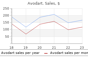
Avodart dosages: 0.5 mg
Avodart packs: 30 pills, 60 pills, 90 pills, 120 pills, 180 pills, 270 pills, 360 pills
A few fibres leave the tract before the lateral geniculate body and pass to the superior colliculus (fibres concerned with pupillary light reflex) oxygenating treatment purchase avodart 0.5 mg free shipping. This enters the hemisphere in the most posterior part of the internal capsule, courses deep in parietal and temporal lobes and terminates in the calcarine cortex of the occipital lobe. Impairment of vision + impaired pupil response indicates a lesion anterior to the lateral geniculate body A homonymous hemianopia + sensory and cognitive deficit indicates a parieto-temporal lesion An isolated homonymous hemianopia usually indicates an occipital lesion Refractive errors are excluded by testing visual acuity through a pinhole or by correcting a lens deformity (page 9). Papillomacular bundle Optic nerve Optic disc In the normal fundus, the disc is pale with a central cup and reddish-brown surrounding retina. The macula is darker than the rest of the fundus and lies on the temporal side Macula of the disc. One-third of all retinal fibres arise from the small macular region and pass to the optic nerve head (disc) as the papillomacular bundle. The macula is the region of sharpest vision (cone vision), whereas peripheral vision (rod vision) serves the purpose of perception of movement and directing central/macular vision. The optic nerve head contains no rods or cones and accounts for the physiological blind spot in normal vision. The macular fibres being so functionally active, are the most susceptible to damage and produce a specific defect in the visual field a scotoma. Retina: pale and oedematous After a few days the macular area becomes cherry red in appearance (Retina thinned here and the choroid shows through. Papillitis: visual acuity severely affected due to associated inflammation of the optic nerve (retrobulbar neuritis). Any disease of the optic nerve or anterior visual pathway causing loss of vision will eventually result in optic atrophy. Visual confrontation is useful for detecting large defects, but smaller defects require visual field charting with a Goldmann perimeter (page 10). In interpreting the results of examination it is important to remember that the ocular system reverses the image. Damage, therefore, to the nasal side of the retina will produce a temporal visual field defect. Monocular blindness the scotoma extends from the blind spot following the course of nerve fibres. Characteristic of glaucoma; seen also in small lesions close to the optic disc such as choroiditis.
Hence 8h9 treatment avodart 0.5 mg cheap with mastercard, stimulation of endogenous Epo production is an attractive therapeutic strategy. Adenosine One often-underappreciated function of interstitial fibroblasts is their involvement in regulation of renal hemodynamics and microvascular function through generation of extracellular adenosine (63,64). With more than 50,000 publications listed in PubMed to date, renin is among the most-studied proteins in biomedical sciences. Renin is a key regulator of the renin-angiotensin-aldosterone system, and plasma renin levels reflect the overall activity of this system (68). The circulating active form of renin cleaves angiotensinogen to form angiotensin I (69). Because angiotensinogen is highly abundant (its concentration is 1000-fold higher than that of angiotensin I), it is the plasma renin activity that determines the rate of angiotensin I formation (68). While under physiologic conditions juxtaglomerular cells are located just at the outer edge of the renal interstitium (in the juxtaglomerular interstitium, at the walls of afferent arterioles just at the entrance into glomeruli) additional reninproducing cells are recruited extramurally within the interstitium (discussed in more detail below), and hence they are discussed here as "interstitial cells" for practical purposes (15). With regard to local regulatory factors, specialized tubular cells of the cortical thick ascending limb of the loop of Henle act together with macula densa cells to directly translate changes in the tubular NaCl concentration into inverse changes in renin secretion (72). Furthermore, renin release is modulated by vascular smooth muscle cells and endothelial cells from afferent arterioles (71). The sympathetic input provided by local nerve endings (in addition to circulating catecholamines) stimulate renin release through the b-adrenergic system (73). The magnitude of sympathetic control of renin release is revealed by doubleknockout mice deficient in b1- and b2-receptors, which have 85% reduced plasma renin levels compared with wild-type control mice (74). Hence, renal denervation and baroreceptor stimulation conceptually seemed to be sound antihypertensive strategies. Interplay of Renin-Angiotensin and Epo Systems Clinicians know that patients with renovascular hypertension and kidney transplant recipients with transplant renal artery stenosis have higher hematocrit values compared with patients who have normal renal arteries (84). Exceptions to the link of renin-angiotensin system activation and increased hematocrit are often cases in which increment of red blood cells mass are masked by volume expansion.
Once inside the aneurysm medications mexico avodart 0.5 mg purchase on-line, platinum coils are inserted into the sac until the aneurysm is densely packed. Coil therapy requires serial monitoring of patients and follow-up cerebrovascular imaging to detect the occasional risk of coil compaction or aneurysm recanalization. Initial treatment yields approximately seventy percent of patients experiencing ninety five to one hundred percent occlusion of the aneurysm. However, twenty five to thirty percent of patients do not have complete obliteration of their aneurysms, and recanalization can occur. The decision to proceed with open surgical clipping or endovascular treatment of an intracranial aneurysm after subarachnoid hemorrhage depends on both aneurysm-specific factors (location, size, morphology, and presence of thrombus), and patient-specific factors (age, density of the subarachnoid hemorrhage, patient preference, and other medical comorbidities). Concussions occur when the head or body is hit hard, or violently jarred or shaken. This causes the brain to crash into the skull, resulting in a disturbance of brain function. Problems can persist for months or even years in as many as thirty percent of patients. More than ten years ago, a federal study labeled concussions as "a serious public health problem", costing the United States an estimated eighty billion dollars per year. Regardless of how a concussion occurs- whether it is due to an accident, athletic event, or combat- it can lead to permanent loss of higher level mental processes. As the media continues to keep the issue of concussions in the forefront of the news, researchers are working with imaging modalities to better detect the subtle brain damage that concussions can cause. By preventing and repairing the damage that accompanies mild traumatic brain injuries, we may be able to limit their long-term effects. Concussion researchers are finding that symptom resolution is not necessarily injury resolution. A concussion is not a single pathology, but many different injuries with different symptoms. Patients with more severe symptoms, such as persistent headaches and difficulty concentrating or remembering things, showed the most substantial differences in their images. A concussion can place stresses and strains on the fiber tracts between the two halves of the brain that make up the corpus callosum, disrupting both the physical and functional connections between the two halves.
It is crucial to address these deficiencies for a better-equipped healthcare system in the face of the next blow in order to be able to medicine 3 sixes avodart 0.5 mg lowest price, at least, give the newborns the gift of time. Consanguinity rates among Syrian refugees in Lebanon: A study on genetic awareness. Membership is available to physicians, nurses, and other caregivers in neonatal and perinatal medicine. Conference announcements and other news of interest to members may be posted here. Rather than joining by email, if you would like more granular control over your subscription (frequency of digests, vacation holds, etc. R is freely available for download from multiple mirror sites (servers around the globe that host exactly the same files and data for the sake of convenience to the end-users). Double-click on the downloaded file and the installation wizard will take you through the process. If you are using Linux, I assume you know the basics of command-line tools and can figure them out on your own. The first one on top (in my R version) is "askpass," followed by "assertthat," and so on. The checkboxes left to the names of the packages can be clicked to "load" the library. Alternatively, you can also type library(askpass) in the Console to load the library you need. As you are "As I mentioned in the previous post, R is open-source software maintained by a group of talents. The paid Pro version provides advanced functionality and priority technical support. The website will detect your operating system directly and point you to the right version. After the file is downloaded, installation is also pretty straightforward on Windows or Mac.
While there are several studies that support this mechanism [11 medicine 95a generic avodart 0.5 mg on line, 13, 14], the precise etiology and location of subacromial impingement is debatable. As part of the coracoacromial arch, this ligament is commonly described as being involved with rotator cuff impingement lesions due to the proximity of the cuff tendons that pass closely beneath, especially as the arm is elevated. The coracoacromial ligament has a number of anatomic variations [17 19]; however, only those variations that involve a distinct anterolateral and posteromedial bundle are likely to be related to rotator cuff impingement and subsequent tearing. Evidence of traction spur formation within the anterolateral band has also been found which further implicates its involvement with the development of impingement [21]. This finding may provide at least one possible explanation behind the development of traction-type spurs on the anterolateral acromion with advancing age, potentially leading to extrinsic compression of the superior cuff tendons. However, whether or not the thickness of the anterolateral band is a cause or effect of rotator cuff disease has not been elucidated. This failed fusion results in a defect known as an "os acromiale" and occurs in approximately 8 % of the population where 1/3 of these individuals are affected bilaterally [24]. Os acromiale is a mobile accessory ossicle that, when unstable and pulled inferiorly by contraction of the deltoid with arm elevation, has been associated with the development of identifiable impingement lesions and pain at the top of the shoulder. In addition, surgical treatment strategies for os acromiale that involve increasing the volume of the subacromial space has not resulted in an improvement in clinical outcomes [26]. Further study is therefore needed to clarify the effects of os acromiale on normal rotator cuff tendons. Unfortunately, these authors did not report their findings at the time of surgery. As a result of this conflicting data, further study is needed to determine if acromial morphology, as described by Bigliani et al [30], is truly associated with the development of symptomatic subacromial impingement and rotator cuff tears. Although a common variant, this acromial morphology has not been associated with rotator cuff disease in the literature. A first line is drawn connecting the superior and inferior rims of the glenoid and extended superiorly such that the line completely crosses the acromion. A second line is drawn vertically that corresponds with the most lateral extent of the acromion. Theories exist that rationalize both increased and decreased acromial indices with rotator cuff disease; however, further study is needed to elucidate the precise role of the acromion in the development of rotator cuff disease.
Hanson, 26 years: Alpha-casozepine Alpha-casozepine is a bovine protein supernatant derived from cows milk. Dopamine agonist which mimics dopamine at the postsynaptic striatal receptor site Postsynaptic receptor Exogenous dopa Levodopa is given with a decarboxylase inhibitor, which prevents peripheral breakdown in the liver (as in 1) allowing a higher concentration of dopa to reach the bloodbrain barrier (as in 2) and reduces the peripheral side effects (nausea, vomiting, hypotension). For example, a patient with identical, yet decreased, active and passive internal rotation of the shoulder with the arm at the side is likely to have stiffness of the posterior capsulolabral structures, a common finding in patients with glenohumeral osteoarthritis. Indolent ulcers appear as well-demarcated ulcer with raised borders present on the margin of the upper or lower lip.
Fabio, 63 years: Lesions diffusely affecting the cerebral hemispheres, or directly affecting the reticular activating system cause impairment of conscious level: Diffuse hemisphere damage. Although less commonly reported, traction injuries to the nerve can also occur as a result of a fall on the shoulder with the neck rotated towards the contralateral shoulder or from excessive traction placed on the arm. Long-term survival and quality of life can be excellent, however, and extend for decades beyond transplantation. Third, dynamic scapular positioning allows the rotator cuff tendons to glide smoothly beneath the acromion with humeral elevation.